
Pet Diagnostic Imaging
At Blue Oasis Veterinary Clinic, we think a correct diagnosis is the initial and most crucial step toward making your pet well again. That’s why we make advanced pet diagnostic imaging a part of our full range of veterinary services. If your pet has a mysterious illness, a suspected injury, or a lingering symptom requiring a closer look, our staff has access to state-of-the-art imaging equipment to determine what’s going on inside the body securely, efficiently, and as painlessly as possible.
Each visit starts with a thorough clinical exam by one of our experienced veterinarians. If what we find at the outset indicates that there’s more at work, we might prescribe imaging as the next step in sorting out your pet’s illness. These non-invasive diagnostic procedures enable us to identify internal abnormalities not visible through a physical exam and arrive at a correct diagnosis sooner, so we can initiate treatment sooner as well.
Our in-house imaging facility has the following:
- Digital X-rays for rapid and precise internal imaging
- Ultrasound equipped with Doppler functionality for evaluation of live organs and circulation
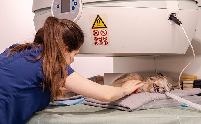
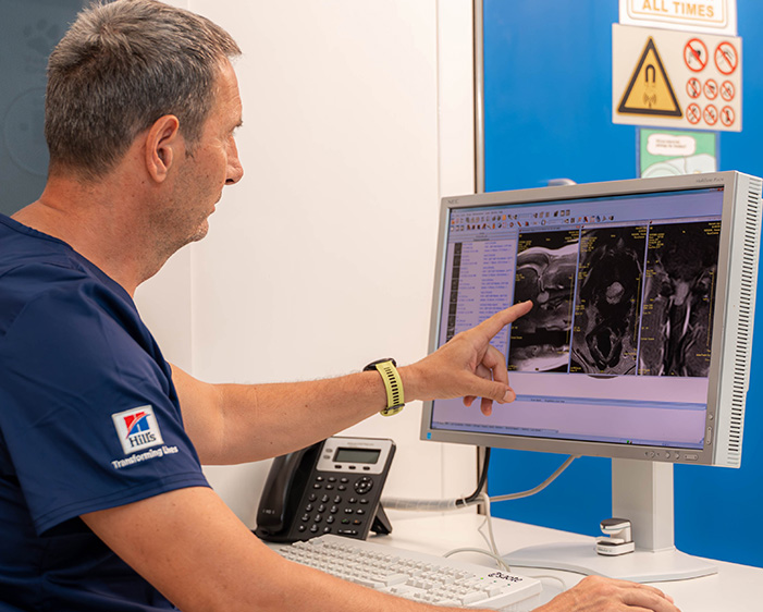
- Magnetic Resonance Imaging (MRI) for high-definition images of the brain, spine and joints
- Endoscopy for internal pathway visualisation and biopsy collection
- ECG for monitoring heart function in real-time
All modalities provide specific benefits and are utilised according to your pet’s specific condition and requirements. At Blue Oasis Veterinary Clinic, we make a judicious decision on the least intrusive yet most informative method so your pet can be given the best possible diagnosis with minimal stress or discomfort.
Why Is Diagnostic Imaging Important?
Diagnostic imaging has a crucial place in contemporary pet medicine. While a physical examination will provide us with a general impression of your pet’s situation on the surface level, imaging allows us to look inside the body and determine internal issues not seen from the outside. Seeing internal organs, tissue, bone, and joints in detail can be the difference between guessing and knowing. For most instances, imaging will be required to:
- Detect bone deformities or fractures.
- Assess the size, shape, and function of organs such as the heart, liver, kidneys, and lungs.
- Diagnose cysts, growths, fluid build-up, or tumours
- Detect gastrointestinal obstructions or foreign objects
- Check the brain and spinal cord for neurological conditions or injuries
- Monitor chronic diseases or monitor healing after surgery
Advanced imaging not only allows us to identify the cause of the problem but also lets us track the evolution of the disease and measure the success of treatment regimens over time. Whether your pet has even the slightest behaviour change or full-blown symptoms, imaging diagnostic can provide key leads for our diagnostic decision-making.
During critical conditions like suspected internal bleeding, trauma, loss of sensitivity, lameness or seizure, imaging allows us to gain life-saving information in real-time and make quick and timely interventions accordingly. At Blue Oasis, we incorporate imaging as part of our holistic approach to animal medicine so that your pet can enjoy the most well-informed, empathetic and tailored care available.
Schedule Your Pet’s Diagnostic Imaging Today
If your pet has a strange symptom, has gotten hurt, or has a long-standing medical issue, don’t wait any longer for the information you’re seeking. The sooner a diagnosis can be made, the sooner we can initiate a customised treatment plan that improves your pet’s comfort level, well-being, and quality of life.
At Blue Oasis Veterinary Clinic, we’re dedicated to making the diagnostic experience easy, transparent, and stress-free for you and your pet. From the moment you step through our doors, our compassionate staff will take you through every step of the way from the initial exam through diagnostic imaging, diagnosis, and follow-up treatment.
Call us today at +971 4 884 8580 to arrange your pet’s diagnostic imaging exam or to consult a member of our team.
Come and see us in Dubai Green Community (DIP 1) and discover for yourself the ways our imaging services can assist in unravelling what’s on the inside of your pet’s body. Your pet deserves answers and Blue Oasis provides them compassionately.
Veterinary x-rays have been in use throughout the medical community for many decades. X-rays are by far the most regularly used form of diagnostic…

An ultrasound is the second most common type of diagnostic imaging tool veterinarians use to diagnose a pet’s medical condition.
Magnetic resonance imaging, or MRI, is the newest form of diagnostic imaging being used for both human and veterinary medicine.
Frequently Asked Questions
Diagnostic Imaging involves the use of medical imaging devices like X-rays, ultrasound, MRI, and other cutting-edge equipment for the diagnosis of diseases, injuries, or abnormalities in any part of the body. At Blue Oasis Veterinary Clinic, our diagnostic imaging procedures are conducted by well-qualified and well-trained veterinary staff utilising the most advanced diagnostic equipment in the UAE for accurate and timely diagnoses of your pet.
Yes. Following your pet’s imaging examination with us, our animal care staff will produce a full diagnostic report summarising the results. In addition, we offer electronic copies of the images (for instance, X-rays, MRI pictures or ultrasound photos) for your keeping. These may be passed on to referring specialists or utilised for future follow-up as required.
This will vary based on the imaging and your pet’s personality. X-rays and ultrasounds usually do not involve sedation unless your pet has pain or anxiety. More involved procedures, such as MRI or endoscopy, have to be performed under anaesthesia so that your pet remains calm and still and out of harm’s way during the scan. We will explain the plan beforehand and prioritise your pet’s comfort and safety throughout the experience.
The amount of time will depend on the type of imaging. As an X-ray will only take a few minutes, an ultrasound scan may take 20 to 30 minutes long, depending on the complexity of the exam, whereas MRIs will take an hour to 90 minutes. If an anaesthesia is necessary, please consider the time for recovery as well. The staff will provide you with an estimate at the time of scheduling and will keep you updated throughout each step of the process.